Difference between revisions of "2024 DOCK tutorial 1 with PDBID 2ITO"
Stonybrook (talk | contribs) (→Fixed Anchor Docking) |
Stonybrook (talk | contribs) (→Virtual Screening of a Ligand Library) |
||
| Line 278: | Line 278: | ||
= Virtual Screening of a Ligand Library = | = Virtual Screening of a Ligand Library = | ||
| + | After we have tested our control case for multiple | ||
=Cartesian Minimization of Virtually Screened Small Molecules= | =Cartesian Minimization of Virtually Screened Small Molecules= | ||
=Rescoring and Ranking Virtually Screened Molecules= | =Rescoring and Ranking Virtually Screened Molecules= | ||
Revision as of 12:39, 12 March 2024
Contents
- 1 Introduction
- 2 Preparation of the ligand and protein
- 3 Creating the Protein Binding Site Surface
- 4 Box and Grid Generation
- 5 Energy Minimization
- 6 DOCK
- 7 Virtual Screening of a Ligand Library
- 8 Cartesian Minimization of Virtually Screened Small Molecules
- 9 Rescoring and Ranking Virtually Screened Molecules
Introduction
For Numbered list do this:
- S
- D
- D
- P
- P
- F
These website tutorials are useful if you are unfamiliar with the following tools:
This tutorial is for PDBID 2ITO. Replace any reference to 2ITO with the protein of your choice.
Learning Objectives
These are for bullets:
- U
- G
Setting Up Your Environment
Sample picture from last year:
Downloading a protein from the PDB database
Preparation of the ligand and protein
Evaluating the Structure
Preparing the Protein file
Preparing the Ligand File
Final Steps
Creating the Protein Binding Site Surface
Creating the Required Surface (DMS) File
Generating Spheres for the Binding Site
Binding Site Spheres
Box and Grid Generation
Generating the Box
T
Generating the Grid
N
Energy Minimization
At
Ligand Minimization
Footprint Analysis
DOCK
Rigid Docking
W
Fixed Anchor Docking
In this session, we will be showing the tutorial for Fixed Anchor Docking. For this mode of docking, the algorithm would first identify the largest fragment extracted from original ligand input. Noted that, the fragments are segmented based on the rotatable bonds of the ligands. Starting from this fragment pose, the algorithm will use it as an anchor to continuously add segments until the whole original ligand pose has been fully re-grown. Noted that when each fragment is added, it would be oriented for a pose that is energetically minimal. Comparing this method to the previous rigid docking mode, fixed anchor docking offer flexibility to the side chain while still being able to keep the general location of the ligand’s binding site as it fixes the anchor position.
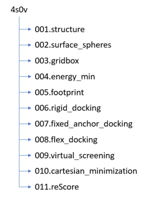
Please navigate to the directory designated for this docking mode: 007.fixed_anchor_docking. Here, we can start create the input parameters, file name – fixed.in:
vi fixed.in
The following is the input parameter for fixed anchor docking. While you can copy and paste this into the body of your script, please make sure to change the input files directory (ligand and protein structure folder) to your personal directory. Also it should be a good practice to make sure the directories for the dock parameters should also be up-to-date.
 conformer_search_type                                        flex
 write_fragment_libraries                                     no
 user_specified_anchor                                        no
 limit_max_anchors                                            no
 min_anchor_size                                              5
 pruning_use_clustering                                       yes
 pruning_max_orients                                          1000
 pruning_clustering_cutoff                                    100
 pruning_conformer_score_cutoff                               100.0
 pruning_conformer_score_scaling_factor                       1.0
 use_clash_overlap                                            no
 write_growth_tree                                            no
 use_internal_energy                                          yes
 internal_energy_rep_exp                                      12
 internal_energy_cutoff                                       100.0
 ligand_atom_file                                             ../004.energy_min/2tio.lig.min_scored.mol2
 limit_max_ligands                                            no
 skip_molecule                                                no
 read_mol_solvation                                           no
 calculate_rmsd                                               yes
 use_rmsd_reference_mol                                       yes
 rmsd_reference_filename                                      ../004.energy_min/2tio.lig.min_scored.mol2
 use_database_filter                                          no
 orient_ligand                                                no
 bump_filter                                                  no
 score_molecules                                              yes
 contact_score_primary                                        no
 contact_score_secondary                                      no
 grid_score_primary                                           yes
 grid_score_secondary                                         no
 grid_score_rep_rad_scale                                     1
 grid_score_vdw_scale                                         1
 grid_score_es_scale                                          1
 grid_score_grid_prefix                                       ../003.gridbox/grid
 multigrid_score_secondary                                    no
 dock3.5_score_secondary                                      no
 continuous_score_secondary                                   no
 footprint_similarity_score_secondary                         no
 pharmacophore_score_secondary                                no
 descriptor_score_secondary                                   no
 gbsa_zou_score_secondary                                     no
 gbsa_hawkins_score_secondary                                 no
 SASA_score_secondary                                         no
 amber_score_secondary                                        no
 minimize_ligand                                              yes
 minimize_anchor                                              yes
 minimize_flexible_growth                                     yes
 use_advanced_simplex_parameters                              no
 simplex_max_cycles                                           1
 simplex_score_converge                                       0.1
 simplex_cycle_converge                                       1
 simplex_trans_step                                           1
 simplex_rot_step                                             0.1
 simplex_tors_step                                            10.0
 simplex_anchor_max_iterations                                500
 simplex_grow_max_iterations                                  500
 simplex_grow_tors_premin_iterations                          0
 simplex_random_seed                                          0
 simplex_restraint_min                                        no
 atom_model                                                   all
 vdw_defn_file                                                /gpfs/projects/AMS536/zzz.programs/dock6.10/parameters/vdw_AMBER_parm99.defn
 flex_defn_file                                               /gpfs/projects/AMS536/zzz.programs/dock6.10/parameters/flex.defn
 flex_drive_file                                              /gpfs/projects/AMS536/zzz.programs/dock6.10/parameters/flex_drive.tbl
 ligand_outfile_prefix                                        2tio_fixed_output
 write_orientations                                           no
 num_scored_conformers                                        1
 rank_ligands                                                 no
Now, you are ready to run dock6 implementing the above input parameters:
dock6 -i fixed.in -o fixed.out
Dock6 should be running for a couple of minutes. If the program finished right away, it would be recommended that you should check the output file to see if there are any reported errors. Successful run should give you two additional files as followed:
- fixed.out
- 2tio_fixed_output_scored.mol2
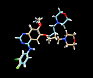
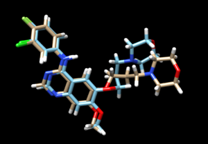
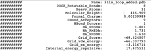
The mol2 file containing the docked ligand could be viewed in ChimeraX under ViewDock. To compare with the reference pose, this mol2 file (blue) has been overlaid on top of the original ligand pose (gold). For this result, it can be seen that although the docking score for the docked ligand is comparable with other docking modes, the RMSD computed for the docked result was larger than 2A, which has been reflected by the pose where the sidechain to the right is shifted further away than in original structure.
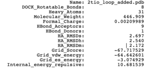
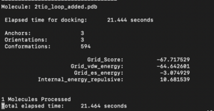
The fixed.out file summary the overall docking score of the run:
The steps for the next docking mode should also be similar.
Fixed Anchor Docking
In this session, we will be showing the tutorial for Flexible Docking. Among the three docking methods introduced in this tutorial, this would be the most computationally expensive method since the algorithm will be sampling all of the possible conformations of each input ligand and test their binding ability with the binding site. Thus, one might expect the computing time for this method to be exceed other introduced methods.
Please navigate to the directory designated for this docking mode: 008.flex_docking. Here, we can start create the input parameters, file name – flex.in:
vi flex.in
The following is the input parameter for fixed anchor docking. While you can copy and paste this into the body of your script, please make sure to change the input files directory (ligand and protein structure folder) to your personal directory. Also it should be a good practice to make sure the directories for the dock parameters should also be up-to-date.
  conformer_search_type                                        flex
 write_fragment_libraries                                     no
 user_specified_anchor                                        no
 limit_max_anchors                                            no
 min_anchor_size                                              5
 pruning_use_clustering                                       yes
 pruning_max_orients                                          1000
 pruning_clustering_cutoff                                    100
 pruning_conformer_score_cutoff                               100.0
 pruning_conformer_score_scaling_factor                       1.0
 use_clash_overlap                                            no
 write_growth_tree                                            no
 use_internal_energy                                          yes
 internal_energy_rep_exp                                      12
 internal_energy_cutoff                                       100.0
 ligand_atom_file                                             ../004.energy_min/2tio.lig.min_scored.mol2
 limit_max_ligands                                            no
 skip_molecule                                                no
 read_mol_solvation                                           no
 calculate_rmsd                                               yes
 use_rmsd_reference_mol                                       yes
 rmsd_reference_filename                                      ../004.energy_min/2tio.lig.min_scored.mol2
 use_database_filter                                          no
 orient_ligand                                                yes
 automated_matching                                           yes
 receptor_site_file                                           ../002.surface_spheres/selected_spheres.sph
 max_orientations                                             1000
 critical_points                                              no
 chemical_matching                                            no
 use_ligand_spheres                                           no
 bump_filter                                                  no
 score_molecules                                              yes
 contact_score_primary                                        no
 contact_score_secondary                                      no
 grid_score_primary                                           yes
 grid_score_secondary                                         no
 grid_score_rep_rad_scale                                     1
 grid_score_vdw_scale                                         1
 grid_score_es_scale                                          1
 grid_score_grid_prefix                                       ../003.gridbox/grid
 multigrid_score_secondary                                    no
 dock3.5_score_secondary                                      no
 continuous_score_secondary                                   no
 footprint_similarity_score_secondary                         no
 pharmacophore_score_secondary                                no
 descriptor_score_secondary                                   no
 gbsa_zou_score_secondary                                     no
 gbsa_hawkins_score_secondary                                 no
 SASA_score_secondary                                         no
 amber_score_secondary                                        no
 minimize_ligand                                              yes
 minimize_anchor                                              yes
 minimize_flexible_growth                                     yes
 use_advanced_simplex_parameters                              no
 simplex_max_cycles                                           1
 simplex_score_converge                                       0.1
 simplex_cycle_converge                                       1.0
 simplex_trans_step                                           1.0
 simplex_rot_step                                             0.1
 simplex_tors_step                                            10.0
 simplex_anchor_max_iterations                                500
 simplex_grow_max_iterations                                  500
 simplex_grow_tors_premin_iterations                          0
 simplex_random_seed                                          0
 simplex_restraint_min                                        no
 atom_model                                                   all
 vdw_defn_file                                                /gpfs/projects/AMS536/zzz.programs/dock10/parameters/vdw_AMBER_parm99.defn
 flex_defn_file                                               /gpfs/projects/AMS536/zzz.programs/dock10/parameters/flex.defn
 flex_drive_file                                              /gpfs/projects/AMS536/zzz.programs/dock10/parameters/flex_drive.tbl
 ligand_outfile_prefix                                        flex.out
 write_orientations                                           no
 num_scored_conformers                                        1
 rank_ligands                                                 no
Now, you are ready to run dock6 implementing the above input parameters:
dock6 -i flex.in -o flex.out
Dock6 should be running for a couple of minutes. If the program finished right away, it would be recommended that you should check the output file to see if there are any reported errors. Successful run should give you two additional files as followed:
- flex.out
- 2tio_flex.out_scored.mol2
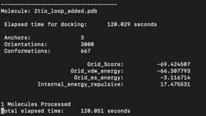
The mol2 file containing the docked ligand could be viewed in ChimeraX under ViewDock. To compare with the reference pose, this mol2 file (blue) has been overlaid on top of the original ligand pose (gold). Under ViewDock in Chimera, it can be observed that the docked ligand showed great structural overlap with the reference pose and this has also been reflected in the reported RMSD score, which has been maintained below 2A.
The fixed.out file summary the overall docking score of the run:
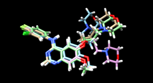
As we have finished running all the docking modes, one step could be carried out here is to compare all the three docked results with the original reference file and see if all the docking results have been suggesting a binding mode that resemble the reference pose. Attached is the overlaid docking results, where we can see the most rigid docking modes give the closest result to the reference structure, whereas fixed anchor docking has the right-side chain shifted further away. Although being the most flexible method, flexible docking gives a good scoring. (gold - original pose, blue - flex, green - rigid, magenta - fixed anchor)
Virtual Screening of a Ligand Library
After we have tested our control case for multiple
