2023 DOCK tutorial 3 with PDBID 2P16
Contents
Introduction
Docking is a molecular modeling program useful for drug discovery. It computes optimal binding conformations and affinities of a small molecule to its target protein using sampling and scoring methods respectively. It enables the discovery of new low-energy ligand binding modes to the active site of a protein, which can help optimize existing drugs or discover new candidates.
DOCK6
DOCK6 was initially developed at UCSF and now is maintained by several groups across three universities. In addition to other features, it introduces new sampling methods such as fixed anchor, de novo design, and genetic algorithms as improvements over traditional rigid docking methods.
In this tutorial we will be exploring the following functions:
- Docking- rigid, flexible, and fixed anchor
- Virtual Screening- docking large libraries of compounds against a protein to generate drug lead
- Cartesian Minimization- energy minimization of docked molecules in the active site
2P16 System
2p16 refers to the pdb code for the crystal structure of Factor Xa in complex with its inhibitor Apixaban.
Factor Xa plays a key role in the formation of blood clots and is therefore a drug target for anticoagulants.
The drug Apixaban is a second-generation therapy developed by Bristol Myers Squibb- it has fewer drug interactions than the more traditional Heparin and Warfarin, and therefore can be used in patients with additional complications. A docking study of this system may elucide the mechanism of this increased specificity and help design the next generation of specific inhibitors.
Directory Structure
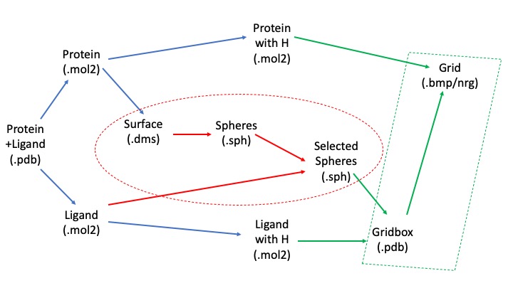
In order to understand the necessary directories it's important to understand the workflow of this project:
We will begin with the pdb file, clean up and separate out the protein and ligand, and then use the unprotonated forms to calculate spheres representing the molecular surface. We will then use our protonated form along with the spheres to calculate the scoring grid. The blue arrows represent transformations we can make using Chimera, the red ones using the SPHGEN and sphere_selector commands, and the green ones using showbox and grid commands.
We will need directories for each step of this process- the sphere generation, grid generation, structure files, as well as for the docking, virtual screening, cartesian minimization, and rescoring after the initial preparatory phase. You can use the following commands to log into the cluster and the generate the relevant directories:
ssh username@login.seawulf.stonybrook.edu
cd /gpfs/projects/AMS536/your_docking_tutorial_directory
mkdir structures spheres gridbox dock virtual_screen virtual_screen_mpi cartesian_min rescore
Protein and Ligand Preparation
Download the 2p16.pdb file from the pdb website, save it in the structures directory, and visualize it using Chimera. You may do so using the command line:
cd structures
wget https://files.rcsb.org/download/2P16.pdb
chimera 2P16.pdb
Make sure you read the associated paper to get an understanding protonation states, charges, and crystallization conditions. Identify the following components- protein, ligand, ions, and solvent.
We will be assuming biological pH and neglecting solvent and ion effects, so you can delete the ions, solvent, and protein chain that isn't involved in ligand binding using the following Chimera commands:
Select->Structure->ion
Actions->Atoms/Bonds->delete

Select->Structure->solvent
Actions->Atoms/Bonds->delete

Select->Chain->L
Actions->Atoms/Bonds->delete
Save the file as 2p16_cleaned.pdb
Protein Preparation
To prepare the protein file delete the ligand:
Select->Structure->ligand
Actions->Atoms/Bonds->delete
Save the file as 2p16_noh_protein.mol2 using File-> Save Mol2
To add hydrogens and charge, you may use the Dockprep function in Chimera through Tools->Surface/Binding Analysis->Dock Prep. Uncheck delete solvent in the first window, as we have done this manually, and press ok until you reach the "Assign Charges" window. Ensure that AMBER ff14SB is selected in this window and AM1-BCC in the following "Specify Net Charges" window. Save the file as 2p16_dockprep_protein.mol2
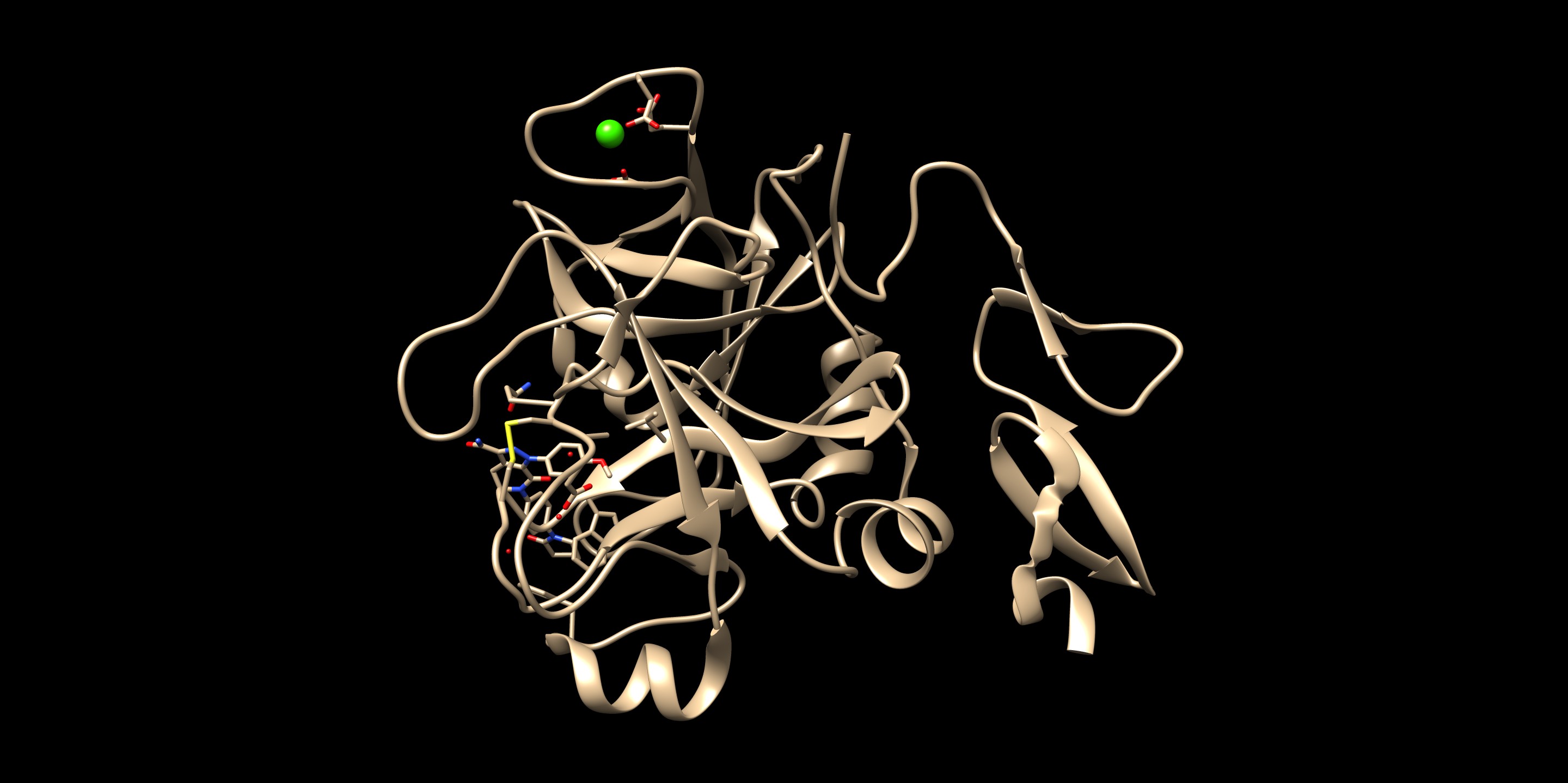
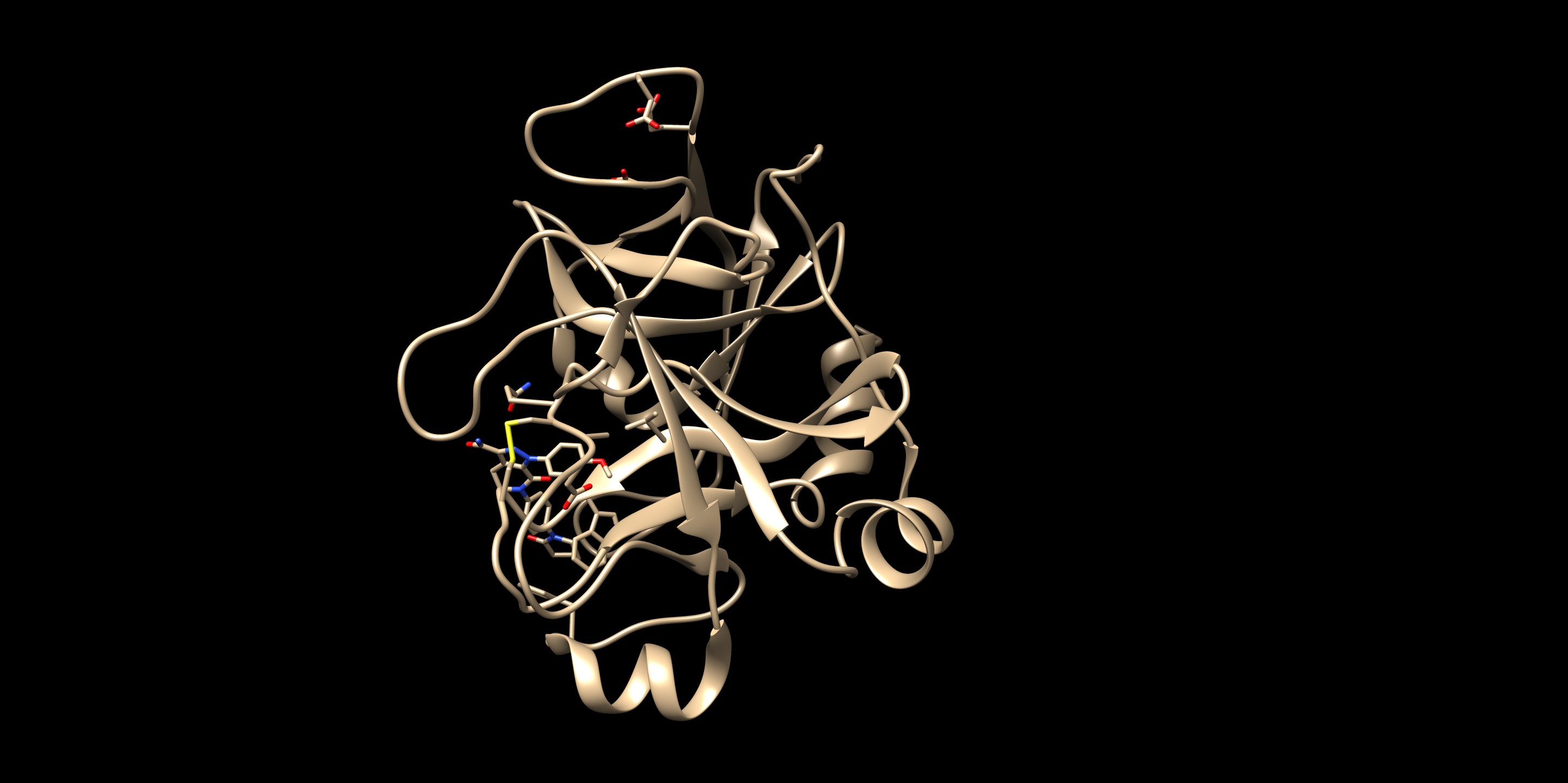
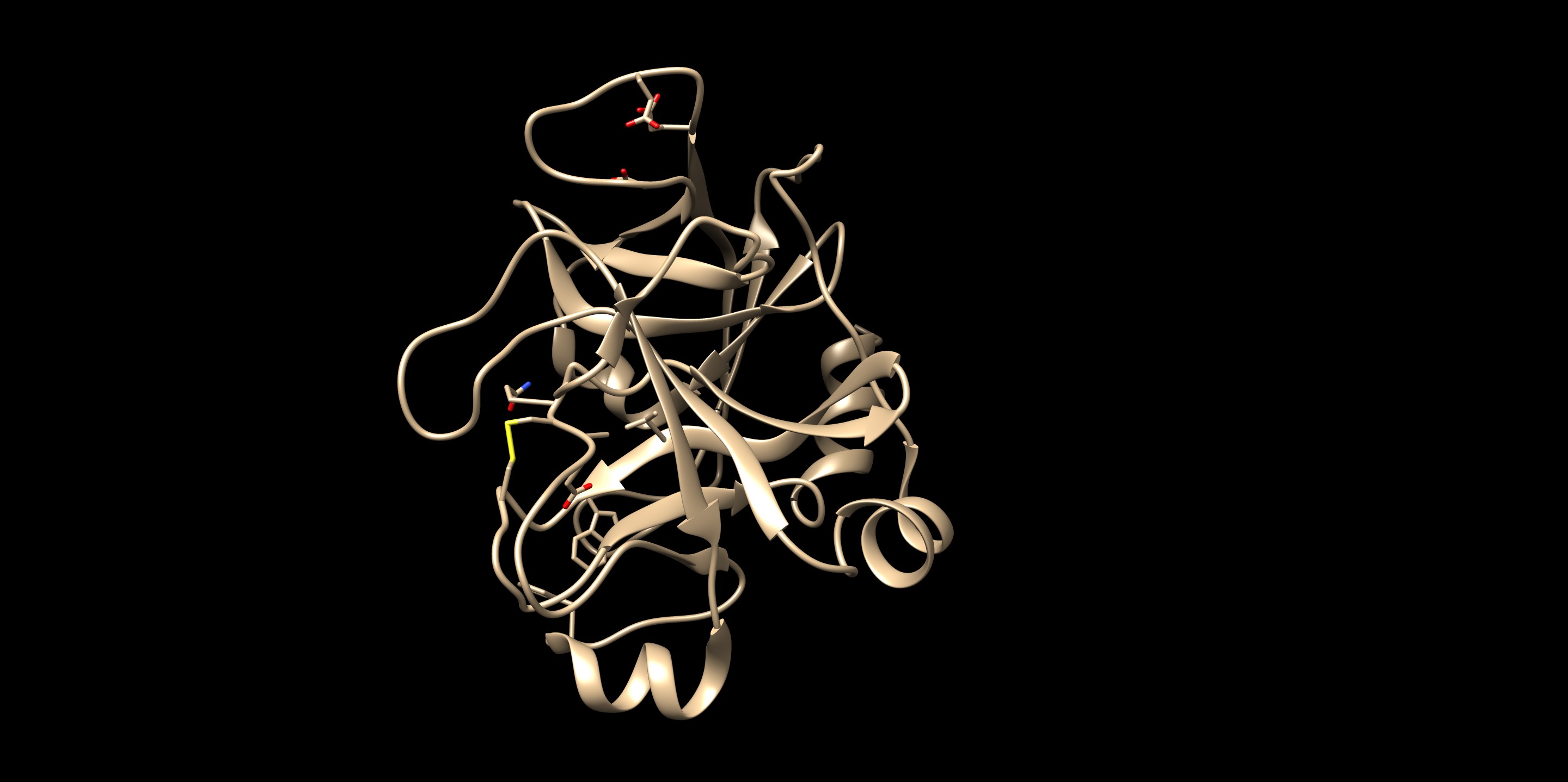
File:2p16 dockprep protein.jpg
Ligand Preparation
To prepare the ligand file load 2p16_cleaned.pdb and delete the protein:
Select->Chain->A
Actions->Atoms/Bonds->delete
Save the file as 2p16_noh_ligand.mol2 using File-> Save Mol2
Add hydrogens and charge using Dockprep. Save the file as 2p16_dockprep_ligand.mol2
Surface Spheres
The remainder of this tutorial will rely on commands from the DOCK6 program. Please ensure that you have DOCK6.10 installed and accessible in your path, which you can check using the commands:
which dock
echo $PATH
If not, you may want to check your ~/.bashrc file and add the following lines if needed:
export DOCKHOME="/gpfs/projects/AMS536/zzz.programs/dock6.10"
 PATH=$DOCKHOME/bin\:$PATH
export PATH=$DOCKHOME/bin\:$PATH
Save the file and activate it using:
source ~/.bashrc
DMS Preparation
To prepare the DMS file open 2p16_noh_protein.mol2 in Chimera and do the following:
Action->Surface->Show
Tools->Structure Editing->Write DMS
Save the file as 2p16_noh_protein.dms
Sphere Generation
Transfer all the files generated so far to the structures directory in your Seawulf docking directory using:
scp 2p16* username@login.seawulf.stonybrook.edu:/gpfs/projects/AMS536/your_docking_tutorial_directory/structures
Move to the spheres directory and open an input file for the sphgen command:
cd spheres
vim INSPH
Type in the following and save the file:
../structures/2p16_noh_protein.dms        #input file
R                                         #spheres to be generated outside the receptor surface
X                                         #all surface points to be used
0.0                                       #surface clashes
4.0.                                      #max sphere radius
1.4.                                      #min sphere radius
2p16_protein.sph                          #output file
Run sphgen using:
sphgen -i INSPH -o OUTSPH
Open your output file 2p16_protein.dms along with your 2016_noh_protein.mol2 file to check if everything looks ok.
Sphere Selection
We now need to narrow down the generated spheres to only those near the binding pocket.
The following command will select those within 10 angstroms of the ligand and save them to selected_spheres.sph:
sphere_selector 2p16_protein.sph ../structures/2p16_noh_ligand.mol2 10.0
File:2p16 selected spheres.jpg
Grid Box
We will now use the selected spheres to generate a grid within which we will score the sampled molecular conformations.
Box Generation
Move to the gridbox directory and open up an input file titled showbox.in. Type in the following text:
Y                                    #Yes to making a box
8.0                                  #Angstroms between box boundary and any sphere
../spheres/selected_spheres.sph      #Input file
1                                    #Number of clusters of spheres to use
2p16.box.pdb                         #Output file
Generate the box using the following command:
showbox < showbox.in
Grid Generation
Open up an input file titled grid.in and type in the following:
compute_grids                             yes
grid_spacing                              0.4
output_molecule                           no
contact_score                             no
energy_score                              yes
energy_cutoff_distance                    9999
atom_model                                a
attractive_exponent                       6
repulsive_exponent                        9
distance_dielectric                       yes
dielectric_factor                         4
bump_filter                               yes
bump_overlap                              0.75
receptor_file                             ../structures/2p16_dockprep_protein.mol2
box_file                                  2p16.box.pdb
vdw_definition_file                       /gpfs/projects/AMS536/zzz.programs/dock6/parameters/vdw_AMBER_parm99.defn
score_grid_prefix                         grid
Generate the grid using the following command:
grid -i grid.in -o grid.out
This should create a grid.out, grid.nrg, and grid.bmp file. Check the grid.out file to see if your residue charges and total charge match the original protein mol2 file. If not, go back and check if you made any mistakes in earlier steps.
Ligand Minimization
Before running DOCK we need to ensure our ligand is energy minimized to prevent any steric clashes. Open up an input file called min.in and type in the following:
conformer_search_type                                        rigid
use_internal_energy                                          yes
internal_energy_rep_exp                                      12
internal_energy_cutoff                                       100.0
ligand_atom_file                                             ../structures/2p16_dockprep_ligand.mol2
limit_max_ligands                                            no
skip_molecule                                                no
read_mol_solvation                                           no
calculate_rmsd                                               yes
use_rmsd_reference_mol                                       ../structures/2p16_dockprep_ligand.mol2
use_database_filter                                          no
orient_ligand                                                no
bump_filter                                                  no
score_molecules                                              yes
contact_score_primary                                        no
contact_score_secondary                                      no
grid_score_primary                                           yes
grid_score_secondary                                         no
grid_score_rep_rad_scale                                     1
grid_score_vdw_scale                                         1
grid_score_es_scale                                          1
grid_score_grid_prefix                                       ../gridbox/grid
multigrid_score_secondary                                    no
dock3.5_score_secondary                                      no
continuous_score_secondary                                   no
footprint_similarity_score_secondary                         no
pharmacophore_score_secondary                                no
descriptor_score_secondary                                   no
gbsa_zou_score_secondary                                     no
gbsa_hawkins_score_secondary                                 no
SASA_score_secondary                                         no
amber_score_secondary                                        no
minimize_ligand                                              yes
simplex_max_iterations                                       1000
simplex_tors_premin_iterations                               0
simplex_max_cycles                                           1
simplex_score_converge                                       0.1
simplex_cycle_converge                                       1.0
simplex_trans_step                                           1.0
simplex_rot_step                                             0.1
simplex_tors_step                                            10.0
simplex_random_seed                                          0
simplex_restraint_min                                        yes
simplex_coefficient_restraint                                10.0
atom_model                                                   all
vdw_defn_file                                                /gpfs/projects/AMS536/zzz.programs/dock6/parameters/vdw_AMBER_parm99.defn
flex_defn_file                                               /gpfs/projects/AMS536/zzz.programs/dock6/parameters/flex.defn
flex_drive_file                                              /gpfs/projects/AMS536/zzz.programs/dock6/parameters/flex_drive.tbl
ligand_outfile_prefix                                        2p16_ligand.min
write_orientations                                           no
num_scored_conformers                                        1
rank_ligands                                                 no
Run the minimization using:
dock6 -i min.in -o min.out
This should save a new file called 2p16_ligand.min_scored.mol2. Open it up in Chimera along with the protein and ligand mol2 files for comparison.
File:2p16 ligand min scored.jpg
You can also view the RMSD values in the header of the file using:
head 2p16.ligand.min_scored.mol2
Footprint Analysis
DOCKING
The goal of docking in this tutorial is to reproduce the crystal structure from x-ray diffraction. This is called “pose reproduction”; this can be used to validate the docking software and parameters of use within the software. If the pose reproduction is not successful, it might be possible that: 1.the parameters to dock the ligand needs to be changed, 2.preparation of ligand or receptor went wrong, 3.the system itself is not suitable to reproduce in the software. Having a preliminary docking pose before getting into virtual screening–a procedure docking thousands of molecules–would be helpful to validate the results you will get in the future. In DOCK6 (at the point of 2020), the docking is successful if the RMSD of the docked pose is within 2.0Å from the crystal pose. In this section, we will be working on (i)rigid docking, (ii)flexible docking, (iii)fixed anchor docking.
Rigid Docking
Rigid docking does not conduct pose searching with dihedral rotations or bond length perturbations. It takes 3 translational and 3 rotational degrees of freedom to do its pose searching. Therefore, rigid docking is less computationally expensive compared to flex docking. Since we already have the crystal structure of the ligand bound at the active site, it is expected to have a good reproduction using rigid docking.
The first step is to create an input file by rigid.in
Insert the following into that file:
conformer_search_type                                        rigid
use_internal_energy                                          yes
internal_energy_rep_exp                                      12
internal_energy_cutoff                                       100
ligand_atom_file                                             2p16.ligand.min_scored.mol2
limit_max_ligands                                            no
skip_molecule                                                no
read_mol_solvation                                           no
calculate_rmsd                                               yes
use_rmsd_reference_mol                                       yes
rmsd_reference_filename                                      2p16.ligand.min_scored.mol2
use_database_filter                                          no
orient_ligand                                                yes
automated_matching                                           yes
receptor_site_file                                           selected_spheres.sph
max_orientations                                             2000
critical_points                                              no
chemical_matching                                            no
use_ligand_spheres                                           no
bump_filter                                                  no
score_molecules                                              yes
contact_score_primary                                        no
contact_score_secondary                                      no
grid_score_primary                                           yes
grid_score_secondary                                         no
grid_score_rep_rad_scale                                     1
grid_score_vdw_scale                                         1
grid_score_es_scale                                          1
grid_score_grid_prefix                                       grid
multigrid_score_secondary                                    no
dock3.5_score_secondary                                      no
continuous_score_secondary                                   no
footprint_similarity_score_secondary                         no
pharmacophore_score_secondary                                no
descriptor_score_secondary                                   no
gbsa_zou_score_secondary                                     no
gbsa_hawkins_score_secondary                                 no
SASA_score_secondary                                         no
amber_score_secondary                                        no
minimize_ligand                                              yes
simplex_max_iterations                                       1000
simplex_tors_premin_iterations                               0
simplex_max_cycles                                           1
simplex_score_converge                                       0.1
simplex_cycle_converge                                       1.0
simplex_trans_step                                           1.0
simplex_rot_step                                             0.1
simplex_tors_step                                            10.0
simplex_random_seed                                          0
simplex_restraint_min                                        no
atom_model                                                   all
vdw_defn_file
/gpfs/projects/AMS536/zzz.programs/dock6.10/parameters/vdw_AMBER_parm99.defn
flex_defn_file
/gpfs/projects/AMS536/zzz.programs/dock6.10/parameters/flex.defn
flex_drive_file
/gpfs/projects/AMS536/zzz.programs/dock6.10/parameters/flex_drive.tbl
ligand_outfile_prefix                                        rigid.out
write_orientations                                           no
num_scored_conformers                                        1
rank_ligands                                                 no
Make sure to (i)have all the files you need (selected_spheres.sph and 2p16.ligand.min_scored.mol2) and (ii)inputs are correct.
Run the input file with the following command:
dock6 -i rigid.in -o rigid.out
After the job is complete, you should see a file named “rigid.out_scored.mol2”. This is the output that shows the docked poses (if you rank it, it will show from the highest to lowest docking score). If you open the file, in the first part, you will see general information about the docked pose including the grid scores. Make sure that the output file is not empty.
The pose can be visualized using Chimera.
After you load the protein you prepared (simply open the file), you can go to:
Tools -> Surface/Binding Analysis -> ViewDock -> *choose the file* -> Dock4,5, or 6.
Along with the visualized pose, you will see a small window popping out with the quantitative information as you saw in the first part of the output file.
Since we took a crystal structure of the ligand, minimized, and rigidly docked into the same protein, it is expected that the produced pose is well-reproduced.
If you want to check the reproducibility, you could check out the RMSD (root-mean-square-deviation) values for the pose.
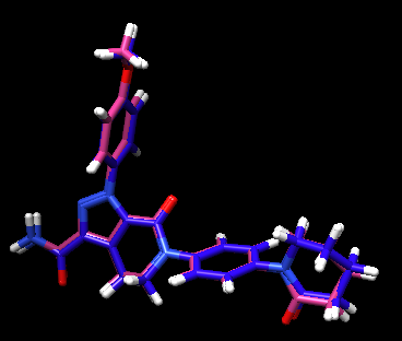
The crystal reference ligand is colored in pink
From the result pose, you can see that the pose reproduction was not successful. From the scored.mol2 file, the RMSD was indicated to be 7.4 Å, which is above 2.0 Å cutoff for a successful pose reproduction. Although this was an unexpected results from the initial assumption, but we can try using two other methods: flex and fixed anchor docking.
Fixed Anchor Docking
As it is said “fixed anchor” docking, this is a docking method that docks while maintaining the orientation of the initially placed structure. For this reason, “comformer_search_type” is flexible, while “write orientation” is turned off. This allows more flexibility in its conformational change compared to rigid docking methods while maintaining the general placement. You can also pick specific fragments within the molecules to be the anchor.
Not to mess up the outputs with other types of docking methods, make a new folder by
mkdir anchor_dock
In this directory, create an input file:
vi fixed.in
Insert the following:
conformer_search_type                                        flex
write_fragment_libraries                                     no
user_specified_anchor                                        no
limit_max_anchors                                            no
min_anchor_size                                              5
pruning_use_clustering                                       yes
pruning_max_orients                                          10000
pruning_clustering_cutoff                                    100
pruning_conformer_score_cutoff                               100.0
pruning_conformer_score_scaling_factor                       1.0
use_clash_overlap                                            no
write_growth_tree                                            no
use_internal_energy                                          yes
internal_energy_rep_exp                                      12
internal_energy_cutoff                                       100.0
ligand_atom_file                                             2p16.charge.mol2
limit_max_ligands                                            no
skip_molecule                                                no
read_mol_solvation                                           no
calculate_rmsd                                               yes
use_rmsd_reference_mol                                       yes
rmsd_reference_filename                                      2p16.charge.mol2
use_database_filter                                          no
orient_ligand                                                no
bump_filter                                                  no
score_molecules                                              yes
contact_score_primary                                        no
contact_score_secondary                                      no
grid_score_primary                                           yes
grid_score_secondary                                         no
grid_score_rep_rad_scale                                     1
grid_score_vdw_scale                                         1
grid_score_es_scale                                          1
grid_score_grid_prefix                                       grid
multigrid_score_secondary                                    no
dock3.5_score_secondary                                      no
continuous_score_secondary                                   no
footprint_similarity_score_secondary                         no
pharmacophore_score_secondary                                no
descriptor_score_secondary                                   no
gbsa_zou_score_secondary                                     no
gbsa_hawkins_score_secondary                                 no
SASA_score_secondary                                         no
amber_score_secondary                                        no
minimize_ligand                                              yes
minimize_anchor                                              yes
minimize_flexible_growth                                     yes
use_advanced_simplex_parameters                              no
simplex_max_cycles                                           1
simplex_score_converge                                       0.1
simplex_cycle_converge                                       1
simplex_trans_step                                           1
simplex_rot_step                                             0.1
simplex_tors_step                                            10
simplex_anchor_max_iterations                                500
simplex_grow_max_iterations                                  500
simplex_grow_tors_premin_iterations                          0
simplex_random_seed                                          0
simplex_restraint_min                                        no
atom_model                                                   all
vdw_defn_file
/gpfs/projects/AMS536/zzz.programs/dock6.10/parameters/vdw_AMBER_parm99.defn
flex_defn_file
/gpfs/projects/AMS536/zzz.programs/dock6.10/parameters/flex.defn
flex_drive_file
/gpfs/projects/AMS536/zzz.programs/dock6.10/parameters/flex_drive.tbl
ligand_outfile_prefix                                        2P16_fixed
write_orientations                                           no
num_scored_conformers                                        1
rank_ligands                                                 no
Start the docking by:
dock6 -i fixed.in -o fixed.out
The resulting docked pose:
The RMSD in this result was 0.22 Å, which is well within 2.0 Å (success cutoff). From the rigid docking result, we could see that the orientation of the entire molecule was flipped from the minimized crystal structure. By having a part of the molecule anchored, it seemed that the big rotation of this molecule did not occur. For a case of new compounds, ones that do not have a crystal structures, this method allows more chemical exploration.
Flex Docking
Finally, flex docking is a docking method that allows flexibility in the rotatable bonds in the ligand and orientation of the compound. This method has the largest exploration of the pose for the ligand, although the calculation time takes the longest. To speed up the calculation time, it is necessary to use cpus provided in seawulf. To use the cpus, we need to submit our run to the queue.
Create a directory:
mkdir flex_dock
First, start with creating an input file, flex.in.
conformer_search_type                                        flex
write_fragment_libraries                                     no
user_specified_anchor                                        no
limit_max_anchors                                            no
min_anchor_size                                              5
pruning_use_clustering                                       yes
pruning_max_orients                                          5000
pruning_clustering_cutoff                                    2500
pruning_conformer_score_cutoff                               100.0
pruning_conformer_score_scaling_factor                       1.0
use_clash_overlap                                            no
write_growth_tree                                            no
use_internal_energy                                          yes
internal_energy_rep_exp                                      12
internal_energy_cutoff                                       100.0
ligand_atom_file
../Rigid_dock/2p16.ligand.min_scored.mol2
limit_max_ligands                                            no
skip_molecule                                                no
read_mol_solvation                                           no
calculate_rmsd                                               yes
use_rmsd_reference_mol                                       yes
rmsd_reference_filename
../Rigid_dock/2p16.ligand.min_scored.mol2
use_database_filter                                          no
orient_ligand                                                yes
automated_matching                                           yes
receptor_site_file                                           ../Rigid_dock/selected_spheres.sph
max_orientations                                             1000
critical_points                                              no
chemical_matching                                            no
use_ligand_spheres                                           no
bump_filter                                                  no
score_molecules                                              yes
contact_score_primary                                        no
contact_score_secondary                                      no
grid_score_primary                                           yes
grid_score_secondary                                         no
grid_score_rep_rad_scale                                     1
grid_score_vdw_scale                                         1
grid_score_es_scale                                          1
grid_score_grid_prefix                                       ../Rigid_dock/grid
multigrid_score_secondary                                    no
dock3.5_score_secondary                                      no
continuous_score_secondary                                   no
footprint_similarity_score_secondary                         no
pharmacophore_score_secondary                                no
descriptor_score_secondary                                   no
gbsa_zou_score_secondary                                     no
gbsa_hawkins_score_secondary                                 no
SASA_score_secondary                                         no
amber_score_secondary                                        no
minimize_ligand                                              yes
minimize_anchor                                              yes
minimize_flexible_growth                                     yes
use_advanced_simplex_parameters                              no
simplex_max_cycles                                           1
simplex_score_converge                                       0.1
simplex_cycle_converge                                       1.0
simplex_trans_step                                           1.0
simplex_rot_step                                             0.1
simplex_tors_step                                            10.0
simplex_anchor_max_iterations                                500
simplex_grow_max_iterations                                  500
simplex_grow_tors_premin_iterations                          0
simplex_random_seed                                          0
simplex_restraint_min                                        no
atom_model                                                   all
vdw_defn_file
/gpfs/projects/AMS536/zzz.programs/dock6.10/parameters/vdw_AMBER_parm99.defn
flex_defn_file
/gpfs/projects/AMS536/zzz.programs/dock6.10/parameters/flex.defn
flex_drive_file
/gpfs/projects/AMS536/zzz.programs/dock6.10/parameters/flex_drive.tbl
ligand_outfile_prefix                                        flex.out
write_orientations                                           yes
num_scored_conformers                                        10
write_conformations                                          yes
cluster_conformations                                        yes
cluster_rmsd_threshold                                       2.0
rank_ligands                                                 no
Now that we have our .in file, we need to make a slurm script to run it. To make a script, first run the following:
vi flex.sh
Inside of this file, copy-paste the following:
 #!/bin/bash
 #SBATCH --time=10:00:00
 #SBATCH --nodes=1
 #SBATCH --ntasks=28
 #SBATCH --job-name=2p16_flex_dock
 #SBATCH --output=2p16_flex_dock
 #SBATCH -p long-28core
dock6 -i flex.in -o 2p16_flex.out
You can submit the queue by
sbatch flex.sh
Once complete, you will see three .mol2 files: (1)flex.out_scored.mol2: best poses scored from the highest to lowest (2)flex.out_conformers.mol2: poses with all conformations of the ligand (3)flex.out_orients.mol2: poses with all orientations of the ligand
To check the best pose, check the flex.out_scored.mol2. You can open it on Chimera using viewdock. Compare with the crystal structure - is the pose with freedom of orientation and rotatable bonds matching with the binding pose in real life?
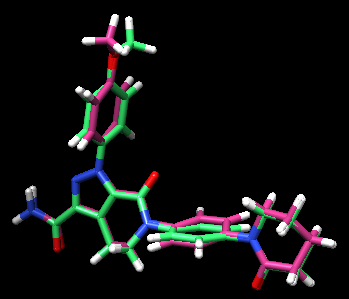
By looking at the comparison (green is the flex output), you can see that the pose reproduction went successfully. The output file showed the RMSD of 0.92 Å, which also indicates a success. This structure was found in the first in scored.mol2 showing that this pose was in fact the best pose after rotational bond and orientation searching.
