File list
This special page shows all uploaded files.
First page |
Previous page |
Next page |
Last page |
| Date | Name | Thumbnail | Size | Description | Versions |
|---|---|---|---|---|---|
| 02:34, 1 March 2020 | 3VJK FLEXIBLE.png (file) |  |
1.71 MB | 1 | |
| 02:31, 1 March 2020 | 3VJK FAD.png (file) |  |
1.67 MB | 1 | |
| 02:12, 1 March 2020 | Rigid entropy.png (file) |  |
1.9 MB | 1 | |
| 23:13, 29 February 2020 | Minimized ligand.png (file) |  |
316 KB | 1 | |
| 15:26, 28 February 2020 | Flex results.jpg (file) | 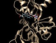 |
447 KB | 1 | |
| 15:26, 28 February 2020 | Fixed results.jpg (file) | 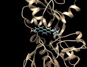 |
444 KB | 1 | |
| 15:23, 28 February 2020 | Rigid results.jpg (file) | 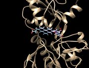 |
446 KB | 1 | |
| 15:11, 28 February 2020 | Footprintpdf.jpeg (file) | 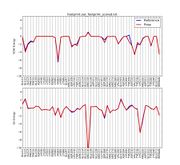 |
485 KB | 1 | |
| 14:30, 26 February 2020 | Flex out.png (file) |  |
276 KB | 1 | |
| 14:15, 26 February 2020 | Fad all.png (file) | 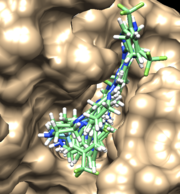 |
456 KB | 1 | |
| 12:42, 26 February 2020 | Fad out.png (file) |  |
325 KB | 2 | |
| 12:39, 26 February 2020 | Rigid all.png (file) | 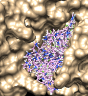 |
283 KB | 2 | |
| 12:30, 26 February 2020 | Rigid out1.png (file) |  |
332 KB | 1 | |
| 16:12, 24 February 2020 | Rigid out.png (file) |  |
650 KB | 1 | |
| 17:53, 20 February 2020 | Footprint output.jpeg (file) | 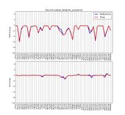 |
816 KB | the figure of the minimized ligand footprint | 1 |
| 17:47, 20 February 2020 | Footprint.jpeg (file) | 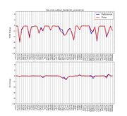 |
457 KB | the figure of footprint | 1 |
| 11:56, 20 February 2020 | 6UZW receptor with the original ligand and the minimized ligand.png (file) |  |
547 KB | 1 | |
| 17:28, 19 February 2020 | Footprint.PNG (file) | 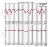 |
46 KB | 1 | |
| 16:15, 19 February 2020 | 4f4p rec noh selected sphere box.png (file) | 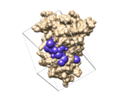 |
872 KB | 4f4p_rec_noh_selected_sphere_box figure | 1 |
| 16:08, 19 February 2020 | Rec min lig complex.png (file) |  |
1.52 MB | 1 | |
| 14:57, 16 February 2020 | 3vjk pdb.png (file) |  |
1.87 MB | 1 | |
| 11:26, 16 February 2020 | 2GQG original.png (file) |  |
335 KB | 1 | |
| 02:21, 16 February 2020 | Sphere 3vjk.png (file) | 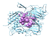 |
358 KB | 1 | |
| 01:59, 16 February 2020 | Sphgen 3vjk.png (file) | 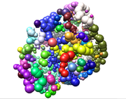 |
459 KB | The spheres represent empty space in the protein structure | 2 |
| 23:19, 15 February 2020 | 3vjk protein.png (file) |  |
894 KB | 1 | |
| 23:02, 15 February 2020 | Ligand with H.png (file) |  |
482 KB | 1 | |
| 22:45, 15 February 2020 | Ligand RAW.png (file) |  |
331 KB | 1 | |
| 22:29, 15 February 2020 | Figure . grid of 6uzw.jpg (file) |  |
160 KB | 1 | |
| 21:16, 15 February 2020 | Grid-image.jpg (file) |  |
160 KB | 1 | |
| 19:40, 15 February 2020 | Surfacev2.png (file) |  |
725 KB | 2 | |
| 19:27, 15 February 2020 | Monomerclean.png (file) |  |
1,007 KB | 2 | |
| 19:21, 15 February 2020 | Monomer20003vjk.png (file) |  |
490 KB | 1 | |
| 18:43, 15 February 2020 | Dimer 3000.png (file) |  |
994 KB | 1 | |
| 16:43, 14 February 2020 | Sphgen 3jvk.png (file) | 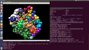 |
846 KB | Results of sphgen. Super temporary photo (hah). For good photos, background should be made white. | 1 |
| 15:29, 13 February 2020 | Lig withH.jpg (file) | 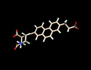 |
118 KB | 1 | |
| 15:28, 13 February 2020 | Lig noH.jpeg (file) | 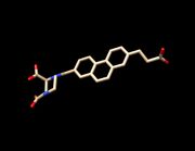 |
93 KB | 1 | |
| 15:26, 13 February 2020 | Lig noH.jpg (file) |  |
93 KB | 1 | |
| 15:24, 13 February 2020 | Selected spheres 6UZW.jpeg (file) | 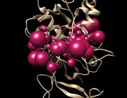 |
258 KB | 1 | |
| 15:21, 13 February 2020 | Spheres 6UZW.jpeg (file) | 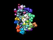 |
221 KB | 3 | |
| 15:16, 13 February 2020 | 6UZW Spheres.jpeg (file) |  |
4 KB | 1 | |
| 15:13, 13 February 2020 | 6UZW Spheres.png (file) |  |
28 KB | 2 | |
| 15:04, 13 February 2020 | Spheres 6UZW.png (file) | 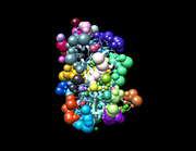 |
751 KB | 1 | |
| 16:48, 12 February 2020 | 4f4pre.png (file) |  |
576 KB | spheres | 1 |
| 16:27, 12 February 2020 | 4f4precnohsph.png (file) |  |
617 KB | spheres | 1 |
| 15:54, 12 February 2020 | 4f4p rec noh surf.png (file) |  |
612 KB | 4f4p_rec_noh_sur | 1 |
| 15:50, 12 February 2020 | 4f4p lig h ams 536.png (file) | 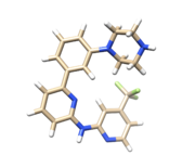 |
434 KB | spring 2020 small molecule for ams 536 class | 1 |
| 18:40, 11 February 2020 | 4f4p lig noh.png (file) |  |
136 KB | 2 | |
| 18:31, 11 February 2020 | 4f4p rec noh.png (file) |  |
596 KB | 2 | |
| 16:38, 11 February 2020 | 4f4p.png (file) |  |
627 KB | Reverted to version as of 15:35, 11 February 2020 (EST) | 3 |
| 12:25, 11 February 2020 | 4F4P.png (file) | 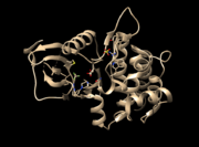 |
185 KB | Figure 1: 4F4P.pdb which contains protein receptor, ligand, sulphate ion and water molecules. (Missing residues in the protein receptor ignored since those are located away from the binding site) | 1 |
First page |
Previous page |
Next page |
Last page |