2019 Covalent docking tutorial 1 with PDB 5VKG
This tutorial teaches you how to dock a covalently bound drug molecule to a receptor (PDB 5VKG) using an alpha version of DOCK6.10.
Use chimera to visualize your system in this case the pdb 5VKG of NMR structure of human Tsg101 UEV in complex with tenatoprazole. PDB 5VKG consists of 20 different models, but we need only one. Either chose one of the models or ask Lauren to give you the file rcsb104645_model1.pdb. Also ask Lauren to provide you with a necessary version of dock6.
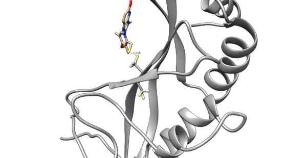
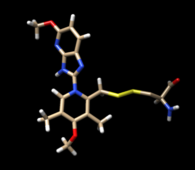
We are interested in the interaction between S atom in Cys 73 and S atom in the ligand. Those two atoms are covalently bonded according to the experimental paper, but Chimera will not show a covalent linker between these two S. We can create the bond ourself in Chimera, if we need (but we don't actually) and measure the distance between these two atoms. If there is a disulfide bond between them, the distance should be 2.05A.
To check the distance we need Shift+Control+left mouse to select two atoms between we want to measure distance.
Tools -> Structure analysis -> Distance.
You will notice the distance from the two sulfurs is about 2.03 angstroms.
Contents
I. Receptor Preparation
First step is to prepare the receptor. To do this open up the original pdb of the molecule. To prepare the receptor, you need to cut off the covalent linkers to generate the receptor.
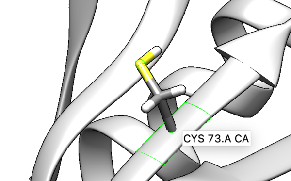
Tools -> Structure editing -> Build Structure -> Modify structure - Element S, Bonds 2, Geometry tetrahedral.
The newly formed receptor is now with a complete cysteine.
Now add hydrogens and charge the protein. To make the cysteine neutral, protons will be added into the system. Protonate with the latest AMBER force field, currently AMBER14SB.
Tools -> Structure editing -> AddH.
Tools -> Structure editing -> AddCharge.
To prepare the receptor for making a grid, delete the side chain of the covalently bonded cysteine residue Cys73. When deleting the side chain of the residue make sure to delete S and the beta carbon, but leave the alpha carbon. That is the receptor that will be used for dock grid and docking.
II. Ligand Preparation
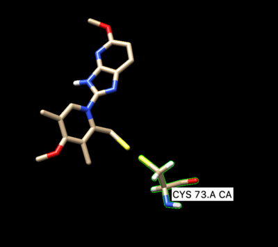
Open of the original structure rcsb104645_model1.pdb. Delete all the protein leaving only the ligand covalently bonded too the cystine, leave the cystine attached to it. The ligand should be binded to the side chain residue all the way up to the beta carbon.
Chose (holding Control + z) Ligand and Cys -> Invert -> Actions -> Atoms -> delete.
Make Cys73 to be visible in all atom mode.
Chose Cys73 (holding Control + z) -> atom/bond -> show.
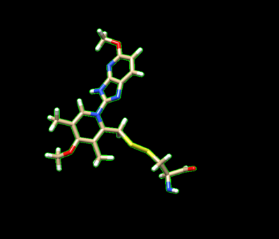
Then we create a disulfide bond between S in the ligand and the residue. The command bellow will create a disulfide covalent bond:
Chose S in the ligand and in the Cys73 (control +z).
Preferences -> Command line -> type "bond sel". It will create a disulfide covalent bond.
Protonate the modified ligand.
Tools -> Structure editing -> AddH
Look at N at the backbone on Cys73 residue. There is only one H attached to N after adding hydrogens, but need to be two. So add one more H to N at the Cys aminoacid backbone. To do so chose N (holding Control +z) and go to
Tools -> Structure editing -> Built structure - Modify Structure - Element N - Bonds 3 - Geometry trigonal.
Next we charge the ligand.
Tools -> Structure editing -> AddCharge -Net charge +1 - with AM1BCC.
Then delete all of the hydrogens off the beta-carbon.
Next we need to modify the beta carbon to D2, and modify the side chain sulfur atom to D1. To do so save prepared ligand in mol2 format and open it with a text editing tool.
Then manually change the atom name of the beta carbon to D2 and change the atom type from C.3 to Du. Change the atom name of the gamma sulfur connected to the beta carbon to D1 and changed the atom type to Du as seen below:
@<TRIPOS>ATOM
     1 D2        -24.0727   34.9285  -36.1952 Du        1 LIG   -0.0660
     2 D1        -25.1580   36.1840  -35.7130 Du        1 LIG   -0.1111
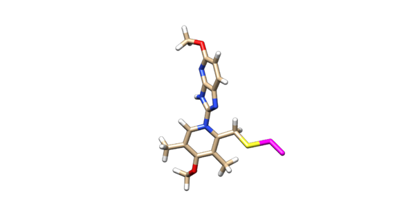
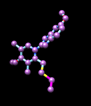
Delete the backbone of the cysteine leaving the beta carbon. The ligand should now look like this in chimera:
This is the end of the ligand preparation.
III. Preparing the sphere files
We need to prepare two types of spheres: 1) spheres for orienting the ligand; 2) spheres for box generating .
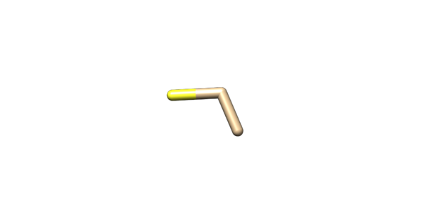
The only spheres that will be orientated will be the alpha carbon, beta carbon, and the sulfur on the cysteine sidechain. Open the mol2 file with prepared ligand. Isolate the sulfur, alpha and beta carbons and delete everything else. Save it into lig_abs.mol2.
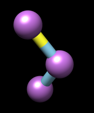
Convert saved coordinates in pdb or mol2 into a sphere file: lig_abs.mol2 lig_abs_spheres.sph
Open file lig_abs_spheres.sph with text editor. Spheres in lig_abs_spheres.sph should be in exact order as 1)S gamma 2)C beta 3)C alpha. Check how atoms are oriented in lig_abs.mol2.
@<TRIPOS>ATOM
     1 CA        -24.6380   34.0060  -37.3390 C.3       1 CYS    0.0000
     2 CB        -24.0170   34.8640  -36.2200 C.3       1 CYS    0.0000
     3 SG        -25.1580   36.1840  -35.7130 S.3       1 CYS    0.0000
It means that in lig_abs_spheres.sph they are also oriented in the same order, but wee need a different order of spheres. Open lig_abs_spheres.sph and rearrange line in correct order by hand.
Now spheres will be generated for the box. A sphere file will be made using the coordinates from the ligand file lig_03_06.mol2. Use only heavy atoms to generate spheres.
IV. Box generating
Now that the spheres are generated twice once for orientating and the other for the dock/grid. The box will be generated. Go to 003.gridbox. Create a file showbox.in. In showbox.in write:
Y
8.0
../002.spheres/lig_spheres.sph
1
lig.box.pdb
Run the command to generate a box:
showbox < showbox.in
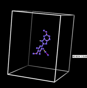
The box result is shown at Figure below.
V. Grid generating
Once the box is made the grid can be populated in the box. Create a file grid.in. But before that copy vdw.defn and chem.defn in current working directory. If don't do this the grid could not recognize the long path for this parameter. Write below into the grid.in file:
compute_grids                             yes
grid_spacing                              0.4
output_molecule                           no
contact_score                             no
energy_score                              yes
energy_cutoff_distance                    9999
atom_model                                a
attractive_exponent                       6
repulsive_exponent                        9
distance_dielectric                       yes
dielectric_factor                         4
bump_filter                               yes
bump_overlap                              0.75
receptor_file                             ../001.files/5VKG_03_06.mol2 change it to ../001.files/5VKG_rec_h.mol2
box_file                                  lig.box.pdb
vdw_definition_file                       vdw.defn
chemical_definition_file               chem.defn
score_grid_prefix                         grid
Run
grid -i grid.in -o grid.out
If this is successful, you will get following output files.
grid.out
grid.nrg
grid.bmp
We can begin covalent docking.
VI. Docking
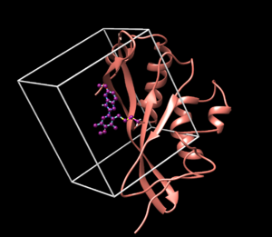
Before starting doing docking open all the prepared structures in chimera to visually observe that everything is prepared correctly. Although when everything looks fine, it still doesn't mean that we prepared everything correctly. But if something looks definitely weird, for example the receptor and the box with a ligand are far from each other, we should think about it and redo.
We can continue with docking. Create a file covalent.in and write the following into it.
conformer_search_type                                        covalent
pruning_use_clustering                                       yes
pruning_max_orients                                          100
pruning_clustering_cutoff                                    100
bondlength                                                   1.8
dihedral_step                                                1.0
pruning_conformer_score_cutoff                               100.0
use_clash_overlap                                            no
write_growth_tree                                            no
use_internal_energy                                          yes
Internal_energy_rep_exp                                      12
internal_energy_cutoff                                       100.0
ligand_atom_file                                             ../001.files/lig_03_06.mol2
limit_max_ligands                                            no
skip_molecule                                                no
read_mol_solvation                                           no
calculate_rmsd                                               yes
rmsd_reference_filename                                      ../001.files/lig_03_06.mol2
use_database_filter                                          no
orient_ligand                                                yes
automated_matching                                           yes
receptor_site_file                                           ../002.spheres/lig_abs_spheres.sph
max_orientations                                             1000
critical_points                                              no
chemical_matching                                            no
use_ligand_spheres                                           no
bump_filter                                                  no
score_molecules                                              yes
contact_score_primary                                        no
grid_score_primary                                           yes
grid_score_rep_rad_scale                                     1
grid_score_vdw_scale                                         1
grid_score_es_scale                                          1
grid_score_grid_prefix                                       ../003.gridbox/grid
minimize_ligand                                              yes
minimize_anchor                                              no
minimize_flexible_growth                                     yes
use_advanced_simplex_parameters                              no
simplex_max_cycles                                           1
simplex_score_converge                                       0.1
simplex_cycle_converge                                       1.0
simplex_trans_step                                           1.0
simplex_rot_step                                             0.1
simplex_tors_step                                            10.0
simplex_grow_max_iterations                                  0
simplex_grow_tors_premin_iterations                          1000
simplex_random_seed                                          0
simplex_restraint_min                                        yes
simplex_coefficient_restraint                                10.0
atom_model                                                   all
vdw_defn_file                                                ../vdw_AMBER_parm99.defn
flex_defn_file                                               ../flex.defn
flex_drive_file                                              ../flex_drive.tbl
ligand_outfile_prefix                                        covalent_out
write_orientations                                           no
num_scored_conformers                                        1000
write_conformations                                          no
cluster_conformations                                        yes
cluster_rmsd_threshold                                       2.0
rank_ligands                                                 no
Run the docking
dock6 -i covalent.in -o covalent.out
To see the output of docking in chimera
Open Chimera
File -> Open -> 5VKG_rec_h.mol2 (Actions -> Surface -> Show)
File -> Open -> lig_03_06.mol2
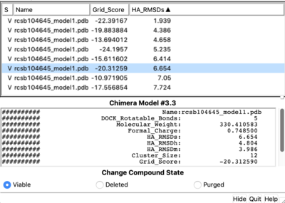
Once everything is loaded go to the ViewDock window and use it's menu to view all the calculated properties regarding the covalent docked ligand by following the steps below.
Tools -> Surface/binding Analysis -> ViewDock -> Select the Covalent Dock output file. (covalent_out_scored.mol2)
 In the loaded dialog box select Dock4,5 or 6
Column -> Show -> Grid_Score
Column -> Show -> HA_RMSDs
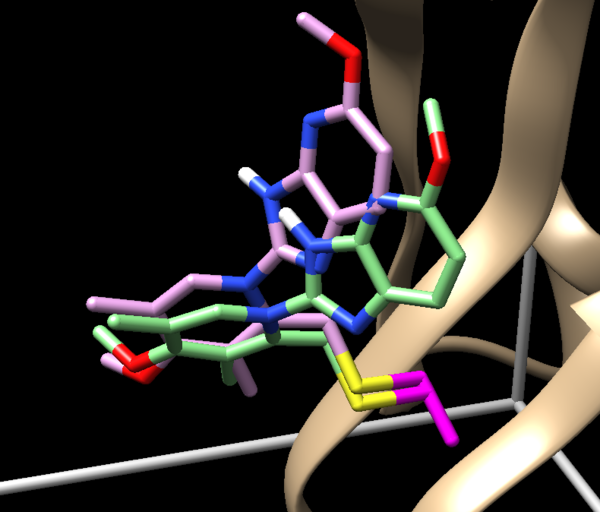
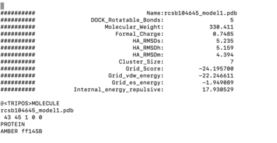
1.9 RMSD is a sampling success. Is it also a scoring success? To understand it we nee to see which of the molecules was ranked first. TO do this let's open covalent_out_scored.mol2 in text editor and see which ligand is appeared first. If it is a molecule with a lowest RMSD than it is a success, but in our case it is not. It means it is a scoring failure. The lowest RMSD structure is ranked second.

