2019 DOCK tutorial 1 with PDBID 2BXF
This tutorial contains a step by step approach to dock a known ligand to a known receptor.
Contents
I. Introduction
DOCK
DOCK is a molecular docking software that was originally developed at UCSF by Dr. Irwin Kuntz, Dr. Brian Shoicket, and colleagues. DOCK is used in drug discovery and molecular modeling. It consists of 2 main components: a search algorithm to explore chemical space for conformations and a scoring function to rank the results.
2BXF
The tutorial will be based on the PDB file 2BXF downloaded from the PDB Database. 2BXF is the crystal structure for human serum albumin complexed with diazepam.
Organization of Directories
We set up the files in our project space as such. It will be helpful to have these folders ready ahead of time.
             0.files
             1.dockprep
             2.surface_spheres
             3.gridbox
             4.dock
             6.footprint
             7.virtual_screen
             8.virtual_screen_mpi
             9.cartesianmin
             10.rescore
II. Preparation of the ligand and receptor
Download the pdb file 2BXF from Protein Data Bank and save the file in the 0.files folder.
Checking the Structure and Preparing the Complex without Hydrogens
- Open Chimera, open the downloaded pdb file, and check the structure for missing residues, gaps, heme groups, missing loops, and size. We recommend using proteins with no heme groups and proteins of a relatively smaller size. 2BXF did not have any of the preceding potential problems.
- Save the PDB file (the ligand and the receptor) as a mol2 file (2BXF_complex_noH.mol2) - When preparing both the ligand the receptor, you can open this complex mol2 file and delete the component that you won't need (eg. for preparing the receptor, open the complex, delete the ligand, and save).
Preparation of Receptor without Hydrogens
 - Open 2BXF_complex_noH.mol2 through Chimera
 - Isolate the receptor by deleting the ligand. Click on the ligand and delete it (Actions -> Atoms/Bonds -> Delete).
 - Save the isolated receptor as a mol2 file (File -> Save mol2 -> 2BXF_rec_noH_mol2)
This receptor has been prepared without Hydrogens.
Preparation of Ligand without Hydrogens
 - Open 2BXF_complex_noH.mol2 through Chimera again
 - Isolate the ligand by deleting the receptor. Click on the receptor and delete it (Actions -> Atoms/Bonds -> Delete).
 - Save the isolated ligand as a mol2 file (File -> Save mol2 -> 2BXF_lig_noH_mol2)
This ligand has been prepared without Hydrogens.
Preparation of receptor with Hydrogens
Preparation of ligand with Hydrogens
-Open the 2BXF_lig_noH.mol2 -Tools -> Surface Binding Analysis
          Tools -> Structure Editing -> Add H (To add Hydrogen atoms)
          Tools -> Structure Editing -> Add Charge (To add the charge use the latest AMBER force filed available for standard residues. Here we used AMBER ff14SB)
          Save as a mol2 file. (2nnq_rec_withH.mol2)
 - If you follow the step below all the above stated steps will automatically appear one after the other to prepare the receptor.
          Tools -> Structure/Binding Analysis -> DockPrep
Preparation of ligand
 - Open the PDB file via Chimera.
 - Using Chimera, isolate the ligand, add H atoms, add charge and save it as a mol2 file by following the same steps followed for the receptor.
Once all the files are prepared make sure to save the files in 1.dockprep folder.
III. Generating receptor surface and spheres
Preparation of DMS file
 - Open 2BXF_rec_noH.mol2 using chimera.
 - Action -> Surface -> Show
 - Tools -> Structure Editing -> Write DMS
 - Save the 2BXF_rec_noH.dms into 3.surface_spheres folder
Reopen the file and make sure the surface was generated.
Transfer all the folders created so far to seawulf cluster to be used in DOCK.
Generating spheres
 - Go to 2.surface_spheres folder
 - Create a new input file to create spheres by typing vim INSPH and type the following lines inside the file.
2BXF_rec_noH.dms
R
X
0.0
4.0
1.4
2BXF_rec.sph
The first line 2BXF_rec_noH.dms specifies the input file. R indicates that spheres generated will be outside of the receptor surface. X specifies all the points will be used. 0.0 is the distance in angstroms and it will avoid steric clashes. 4.0 is the maximum surface radius of the spheres and 1.4 is the minimum radius in angstroms.The last line 2BXF_spheres.sph creates the sph file that contains clustered spheres.
Once the INSPH file is ready, type the following command to generate the spheres.
sphgen -i INSPH -o OUTSPH
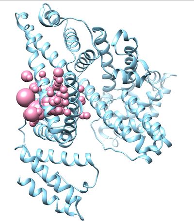
Once sphgen command is successful, 2BXF_spheres.sph file will be created. Open it up using Chimera along with 2BXF_rec_noH.mol2 file. You should get a similar output like the image below.
Selecting Spheres
Here we will be selecting the spheres which defines the binding pocket of the ligand because we are trying to direct the ligand towards that binding site rather than all over the receptor. To select the spheres type the following command.
sphere_selector 2BXF_rec.sph ../1.dockprep/2BXF_lig_withH.mol2 10.0
This command will select all of the spheres within 10.0 angstroms of the ligand and output them to selected_spheres.sph. Visualize the selected spheres using Chimera to make sure the correct spheres are selected. Notice that, spheres around the ligand binding site are kept and all the other spheres are deleted in the image below.
IV. Generating box and grid
Generating box
Move to 3.boxgrid directory Create a new file showbox.in and write the following lines in the file.
 Y
 8.0
 ../2.surface_spheres/selected_spheres.sph
 1
 2BXF.box.pdb
Each of the above lines indicate that;
 We intend to generate a box
 The box length should be 8 Angstroms
 Use the selected_spheres file in the designated location
 The name of the file that contains generated box.
Use the following command to generate the box.
showbox < showbox.in
If this step is successful, you should see a new file (2BXF.box.pdb) in 3.boxgrid folder.
Generating grid
Create a new file (grid.in)
Use the following command to generate the grid.
grid -i grid.in -o gridinfo.out
Answer the prompted questions with the answers given below. (or you can use the following lines and include them in the grid.in file before entering the above command. If you do that these questions won't be prompted again. They will be automatically answered by grid.in file created)
compute_grids                             yes
grid_spacing                              0.4
output_molecule                           no
contact_score                             no
energy_score                              yes
energy_cutoff_distance                    9999
atom_model                                a
attractive_exponent                       6
repulsive_exponent                        9
distance_dielectric                       yes
dielectric_factor                         4
bump_filter                               yes
bump_overlap                              0.75
receptor_file                             ../1.dockprep/2BXF_rec_withH.mol2
box_file                                  2BXF.box.pdb
vdw_definition_file                       /gpfs/projects/AMS536/zzz.programs/dock6/parameters/vdw_AMBER_parm99.defn
score_grid_prefix                         grid
If the command is successful, three new files will be generated. (gridinfo.out, grid.nrg, grid.bmp). Go through gridinfo.out file to make sure all the information about the receptor in the file matches with the original information of the receptor. (Eg:- Total charge, residues and their charges) If the information doesn't match, that means you have made an error in one of the steps that you followed so far.
V. Docking a single molecule for pose reproduction
Under this section, the ligand for 2BXF.pdb will be re-docked into the receptor. 3 Methods will be used to achieve this.
1. rigid docking
2. fixed anchor docking
3. flexible docking
Energy minimization
Before performing docking, here the ligand will be subjected to energy minimization in order to remove unfavorable clashes. These clashes will affect rigid docking because in rigid docking the ligand will be docked as the complete ligand, whereas in other docking methods the ligand will be broken into fragments and the ligand will be built step by step considering favorable orientations and torsion angles after each fragment addition.
Go to the directory 4.dock and a create a new file (min.in) and enter the command below.
dock6 -i min.in -o min.out
Answer the prompted questions using the answers given below or include the following lines in the min.in file at before entering the above command to avoid answering the questions manually.
conformer_search_type                                        rigid
use_internal_energy                                          yes
internal_energy_rep_exp                                      12
internal_energy_cutoff                                       100.0
ligand_atom_file                                             ../1.dockprep/2BXF_lig_withH.mol2
limit_max_ligands                                            no
skip_molecule                                                no
read_mol_solvation                                           no
calculate_rmsd                                               yes
use_rmsd_reference_mol                                 yes
rmsd_reference_filename                                 ../1.dockprep/2BXF_lig_withH.mol2
use_database_filter                                          no
orient_ligand                                                no
bump_filter                                                  no
score_molecules                                              yes
contact_score_primary                                        no
contact_score_secondary                                      no
grid_score_primary                                           yes
grid_score_secondary                                         no
grid_score_rep_rad_scale                                     1
grid_score_vdw_scale                                         1
grid_score_es_scale                                          1
grid_score_grid_prefix                                       ../3.boxgrid/grid
multigrid_score_secondary                                    no
dock3.5_score_secondary                                      no
continuous_score_secondary                                   no
footprint_similarity_score_secondary                         no
pharmacophore_score_secondary                                no
descriptor_score_secondary                                   no
gbsa_zou_score_secondary                                     no
gbsa_hawkins_score_secondary                                 no
SASA_score_secondary                                         no
amber_score_secondary                                        no
minimize_ligand                                              yes
simplex_max_iterations                                       1000
simplex_tors_premin_iterations                               0
simplex_max_cycles                                           1
simplex_score_converge                                       0.1
simplex_cycle_converge                                       1.0
simplex_trans_step                                           1.0
simplex_rot_step                                             0.1
simplex_tors_step                                            10.0
simplex_random_seed                                          0
simplex_restraint_min                                        yes
simplex_coefficient_restraint                                10.0
atom_model                                                   all
vdw_defn_file                                                /gpfs/projects/AMS536/zzz.programs/dock6/parameters/vdw_AMBER_parm99.defn
flex_defn_file                                               /gpfs/projects/AMS536/zzz.programs/dock6/parameters/flex.defn
flex_drive_file                                              /gpfs/projects/AMS536/zzz.programs/dock6/parameters/flex_drive.tbl
ligand_outfile_prefix                                        2BXF.lig.min
write_orientations                                           no
num_scored_conformers                                        1
rank_ligands                                                 no
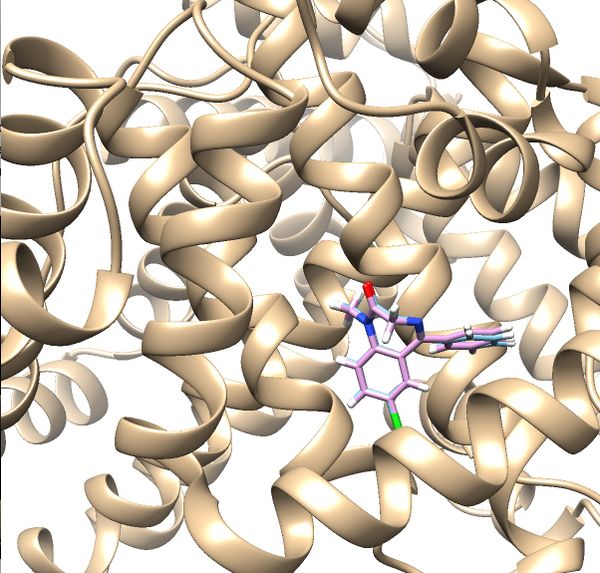
If the process is successful a new file (2BXF.lig.min_scored.mol2) will be generated. You can compare how is it changed from the initial structure by analyzing the RMSD value generated in the file. Visualize the new mol2 file along with receptor and the initial ligand mol2 files using Chimera to see the differences.
Rigid Docking
Create an input file for rigid docking
touch rigid.in
Run dock using the created input file.
dock6 -i rigid.in -o rigid.out
Follow a similar approach as we did for minimization to answer the prompted questions by either answering them manually using the answers in the lines below or by including the following lines in the input file before running dock.
conformer_search_type                                        rigid
use_internal_energy                                          yes
ligand_atom_file                                             2BXF.lig.min_scored.mol2
limit_max_ligands                                            no
skip_molecule                                                no
read_mol_solvation                                           no
calculate_rmsd                                               yes
use_rmsd_reference_mol                                 yes
rmsd_reference_filename                                  2BXF.lig.min_scored.mol2
use_database_filter                                          no
orient_ligand                                                yes
automated_matching                                           yes
receptor_site_file                                           ../2.surface_spheres/selected_spheres.sph
max_orientations                                             1000
critical_points                                              no
chemical_matching                                            no
use_ligand_spheres                                           no
bump_filter                                                  no
score_molecules                                              yes
contact_score_primary                                        no
contact_score_secondary                                      no
grid_score_primary                                           yes
grid_score_secondary                                         no
grid_score_rep_rad_scale                                     1
grid_score_vdw_scale                                         1
grid_score_es_scale                                          1
grid_score_grid_prefix                                       ../3.boxgrid/grid
multigrid_score_secondary                                    no
dock3.5_score_secondary                                      no
continuous_score_secondary                                   no
footprint_similarity_score_secondary                         no
pharmacophore_score_secondary                                no
descriptor_score_secondary                                   no
gbsa_zou_score_secondary                                     no
gbsa_hawkins_score_secondary                                 no
SASA_score_secondary                                         no
amber_score_secondary                                        no
minimize_ligand                                              yes
simplex_max_iterations                                       1000
simplex_tors_premin_iterations                               0
simplex_max_cycles                                           1
simplex_score_converge                                       0.1
simplex_cycle_converge                                       1.0
simplex_trans_step                                           1.0
simplex_rot_step                                             0.1
simplex_tors_step                                            10.0
simplex_random_seed                                          0
simplex_restraint_min                                        no
atom_model                                                   all
vdw_defn_file                                                /gpfs/projects/AMS536/zzz.programs/dock6/parameters/vdw_AMBER_parm99.defn
flex_defn_file                                               /gpfs/projects/AMS536/zzz.programs/dock6/parameters/flex.defn
flex_drive_file                                              /gpfs/projects/AMS536/zzz.programs/dock6/parameters/flex_drive.tbl
ligand_outfile_prefix                                        rigid.out
write_orientations                                           no
num_scored_conformers                                        1
rank_ligands                                                 no
Once rigid docking is successful, you will get an output file. (rigid.out_scored.mol2) Visualize the output file using Chimera by following steps to check the rigid docking success.
Open Chimera
File -> Open -> 2BXF_rec_withH.mol2
File -> Open -> 2BXF_lig_withH.mol2
Tools -> Surface/binding Analysis -> ViewDock -> Select the Rigid Dock output file. (rigid.out_scored.mol2)
In the loaded dialog box select Dock4,5 or 6
Once everything is loaded go to the ViewDock window and use it's menu to view all the calculated properties regarding the rigid docked ligand by following the steps below.
Column -> Show -> gridscore
Column -> Show -> HA_RMSDs
Follow the same steps to get all the properties
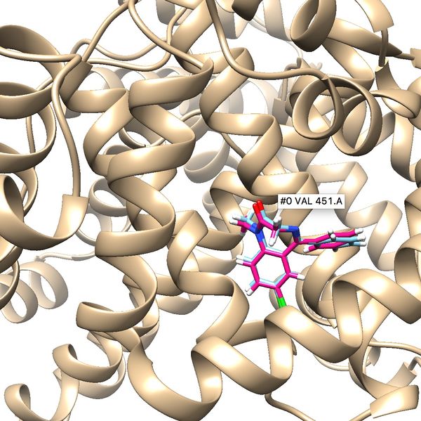
Your visualized structure should be similar to the image below.
Fixed Anchor Docking
Create an input file for fixed anchor docking.
touch fixed.in
Use the input file to perform fixed anchor docking
dock6 -i fixed.in
Use the following lines to answer the prompted questions as we did in rigid docking.
conformer_search_type                                        flex
user_specified_anchor                                        no
limit_max_anchors                                            no
min_anchor_size                                              5
pruning_use_clustering                                       yes
pruning_max_orients                                          1000
pruning_clustering_cutoff                                    100
pruning_conformer_score_cutoff                               100.0
pruning_conformer_score_scaling_factor                       1.0
use_clash_overlap                                            no
write_growth_tree                                            no
write_fragment_libraries                                     no
use_internal_energy                                          yes
internal_energy_rep_exp                                      12
internal_energy_cutoff                                       100.0
ligand_atom_file                                             ../1.dockprep/2BXF_lig_withH.mol2
limit_max_ligands                                            no
skip_molecule                                                no
read_mol_solvation                                           no
calculate_rmsd                                               yes
use_rmsd_reference_mol                                       yes
rmsd_reference_filename                                      ../1.dockprep/2BXF_lig_withH.mol2
use_database_filter                                          no
orient_ligand                                                no
bump_filter                                                  no
score_molecules                                              yes
contact_score_primary                                        no
contact_score_secondary                                      no
grid_score_primary                                           yes
grid_score_secondary                                         no
grid_score_rep_rad_scale                                     1
grid_score_vdw_scale                                         1
grid_score_es_scale                                          1
grid_score_grid_prefix                                       ../2.boxgrid/grid
multigrid_score_secondary                                    no
dock3.5_score_secondary                                      no
continuous_score_secondary                                   no
footprint_similarity_score_secondary                         no
pharmacophore_score_secondary                                no
descriptor_score_secondary                                   no
gbsa_zou_score_secondary                                     no
gbsa_hawkins_score_secondary                                 no
SASA_score_secondary                                         no
amber_score_secondary                                        no
minimize_ligand                                              yes
minimize_anchor                                              yes
minimize_flexible_growth                                     yes
use_advanced_simplex_parameters                              no
simplex_max_cycles                                           1
simplex_score_converge                                       0.1
simplex_cycle_converge                                       1.0
simplex_trans_step                                           1.0
simplex_rot_step                                             0.1
simplex_tors_step                                            10.0
simplex_anchor_max_iterations                                500
simplex_grow_max_iterations                                  500
simplex_grow_tors_premin_iterations                          0
simplex_random_seed                                          0
simplex_restraint_min                                        no
atom_model                                                   all
vdw_defn_file                                                /gpfs/projects/AMS536/zzz.programs/dock6/parameters/vdw_AMBER_parm99.defn
flex_defn_file                                               /gpfs/projects/AMS536/zzz.programs/dock6/parameters/flex.defn
flex_drive_file                                              /gpfs/projects/AMS536/zzz.programs/dock6/parameters/flex_drive.tbl
ligand_outfile_prefix                                        2BXF_fad
write_orientations                                           no
num_scored_conformers                                        100
write_conformations                                          no
cluster_conformations                                        yes
cluster_rmsd_threshold                                       2.0
rank_ligands                                                 no
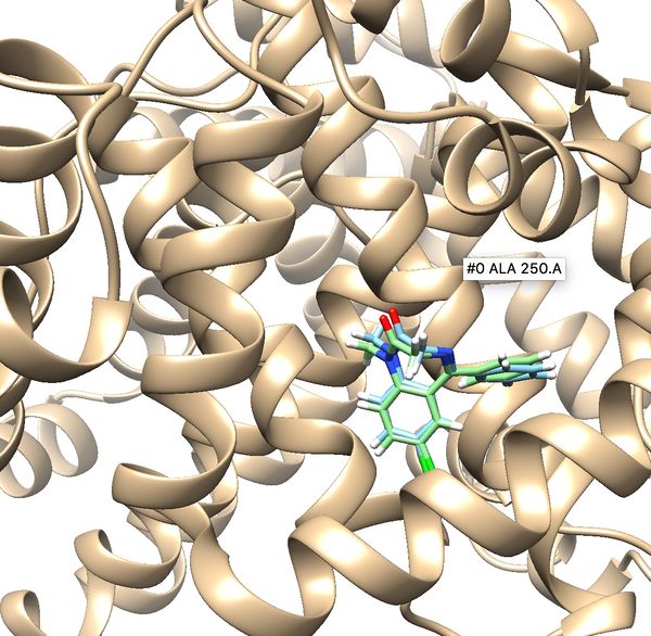
Once docking is completed an output file will be generated. (2BXF_fad_scored.mol2) Follow the same method used in rigid docking to visualize the docked poses using Chimera. Once it is visualized, it should like the image below. Notice all the poses 50 generated are in the same cluster and standard RMSD is 0.75. These indicate that docking is very successful.
Flexible Docking
Create a new input file for flexible docking. (flex.in)
touch flex.in
Use the created input file to perform flexible docking using DOCK6.
dock6 -i flex.in -o flex.out
Answer the prompted questions using the following lines as we did in rigid and fixed anchor docking.
conformer_search_type                                        flex
user_specified_anchor                                        no
limit_max_anchors                                            no
min_anchor_size                                              5
pruning_use_clustering                                       yes
pruning_max_orients                                          1000
pruning_clustering_cutoff                                    100
pruning_conformer_score_cutoff                               100.0
pruning_conformer_score_scaling_factor                       1.0
use_clash_overlap                                            no
write_growth_tree                                            no
write_fragment_libraries                                     no
use_internal_energy                                          yes
internal_energy_rep_exp                                      12
internal_energy_cutoff                                       100.0
ligand_atom_file                                             2BXF.lig.min_scored.mol2
limit_max_ligands                                            no
skip_molecule                                                no
read_mol_solvation                                           no
calculate_rmsd                                               yes
use_rmsd_reference_mol                                yes
rmsd_reference_filename                                2BXF.lig.min_scored.mol2
use_database_filter                                          no
orient_ligand                                                yes
automated_matching                                           yes
receptor_site_file                                           ../2.surface_spheres/selected_spheres.sph
max_orientations                                             1000
critical_points                                              no
chemical_matching                                            no
use_ligand_spheres                                           no
bump_filter                                                  no
score_molecules                                              yes
contact_score_primary                                        no
contact_score_secondary                                      no
grid_score_primary                                           yes
grid_score_secondary                                         no
grid_score_rep_rad_scale                                     1
grid_score_vdw_scale                                         1
grid_score_es_scale                                          1
grid_score_grid_prefix                                       ../3.boxgrid/grid
multigrid_score_secondary                                    no
dock3.5_score_secondary                                      no
continuous_score_secondary                                   no
footprint_similarity_score_secondary                         no
pharmacophore_score_secondary                                no
descriptor_score_secondary                                   no
gbsa_zou_score_secondary                                     no
gbsa_hawkins_score_secondary                                 no
SASA_score_secondary                                         no
amber_score_secondary                                        no
minimize_ligand                                              yes
minimize_anchor                                              yes
minimize_flexible_growth                                     yes
use_advanced_simplex_parameters                              no
simplex_max_cycles                                           1
simplex_score_converge                                       0.1
simplex_cycle_converge                                       1.0
simplex_trans_step                                           1.0
simplex_rot_step                                             0.1
simplex_tors_step                                            10.0
simplex_anchor_max_iterations                                500
simplex_grow_max_iterations                                  500
simplex_grow_tors_premin_iterations                          0
simplex_random_seed                                          0
simplex_restraint_min                                        no
atom_model                                                   all
vdw_defn_file                                                /gpfs/projects/AMS536/zzz.programs/dock6/parameters/vdw_AMBER_parm99.defn
flex_defn_file                                               /gpfs/projects/AMS536/zzz.programs/dock6/parameters/flex.defn
flex_drive_file                                              /gpfs/projects/AMS536/zzz.programs/dock6/parameters/flex_drive.tbl
ligand_outfile_prefix                                        flex.out
write_orientations                                           no
num_scored_conformers                                        1
rank_ligands                                                 no
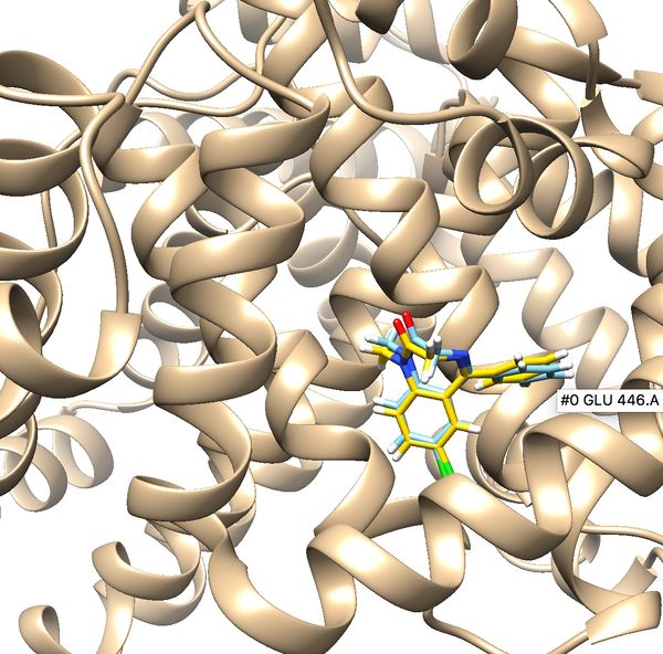
Once flexible docking is completed an output mol2 file will be generated. (flex.out_scored.mol2). Use the visualization steps used in rigid and fixed anchor docking and study the properties of the docking results.
Molecular Footprint
Molecular footprints can be used to determine how a ligand interacts with the receptor. Usually, the molecular footprint shows electrostatic interactions and Van der Waals interactions. Here, the molecular footprint will be used to determine how the ligand interacts with the receptor before and after minimization. To generate molecular footprints use following steps.
Go to directory 6.footprint
Generate an input file by typing;
touch footprint.in
Use DOCK6 to generate footprints
dock6 -i footprint.in
Use the following lines to answer the prompted questions.
conformer_search_type                                        rigid
use_internal_energy                                          no
ligand_atom_file                                             2BXF_lig_min.mol2
limit_max_ligands                                            no
skip_molecule                                                no
read_mol_solvation                                           no
calculate_rmsd                                               no
use_database_filter                                          no
orient_ligand                                                no
bump_filter                                                  no
score_molecules                                              yes
contact_score_primary                                        no
contact_score_secondary                                      no
grid_score_primary                                           no
grid_score_secondary                                         no
multigrid_score_primary                                      no
multigrid_score_secondary                                    no
dock3.5_score_primary                                        no
dock3.5_score_secondary                                      no
continuous_score_primary                                     no
continuous_score_secondary                                   no
footprint_similarity_score_primary                           yes
footprint_similarity_score_secondary                         no
fps_score_use_footprint_reference_mol2                       yes
fps_score_footprint_reference_mol2_filename                  2BXF_lig_with.mol2
fps_score_foot_compare_type                                  Euclidean
fps_score_normalize_foot                                     no
fps_score_foot_comp_all_residue                              yes
fps_score_receptor_filename                                  ../1.dockprep/2BXF_rec_withH.mol2
fps_score_vdw_att_exp                                        6
fps_score_vdw_rep_exp                                        12
fps_score_vdw_rep_rad_scale                                  1
fps_score_use_distance_dependent_dielectric                  yes
fps_score_dielectric                                         4.0
fps_score_vdw_fp_scale                                        1
fps_score_es_fp_scale                                        1
fps_score_hb_fp_scale                                        0
pharmacophore_score_secondary                                no
descriptor_score_secondary                                   no
gbsa_zou_score_secondary                                     no
gbsa_hawkins_score_secondary                                 no
SASA_score_secondary                                         no
amber_score_secondary                                        no
minimize_ligand                                              no
atom_model                                                   all
vdw_defn_file                                                /gpfs/projects/AMS536/zzz.programs/dock6/parameters/vdw_AMBER_parm99.defn
flex_defn_file                                               /gpfs/projects/AMS536/zzz.programs/dock6/parameters/flex.defn
flex_drive_file                                              /gpfs/projects/AMS536/zzz.programs/dock6/parameters/flex_drive.tbl
ligand_outfile_prefix                                        footprint.out
write_footprints                                             yes
write_hbonds                                                 yes
write_orientations                                           no
num_scored_conformers                                        1
rank_ligands                                                 no
Once everything is successful these output files should be generated. (footprint.out_footprint_scored.txt, footprint.out_hbond_scored.txt, footprint.out_scored.mol2)
Use a python script to visualize the molecular footprint. The script can be accessed in the previous DOCK tutorials. Once the script is used, the molecular footprint should be similar to the image below. Notice the large deviations in energy at different amino acid residues. Those residues contribute more towards, the interaction between the ligand and therefore can be identified as important towards the binding of new ligands which can replace the original ligand in the PDB file.

